Journal of South China University of Technology(Natural Science Edition) ›› 2022, Vol. 50 ›› Issue (12): 132-141.doi: 10.12141/j.issn.1000-565X.220042
Special Issue: 2022年电子、通信与自动控制
• Electronics, Communication & Automation Technology • Previous Articles Next Articles
A Multi-Scale Lightweight Brain Glioma Image Segmentation Network
YANG Jinsheng CHEN Hongpeng GUAN Xin LI Qiang
- School of Microelectronics,Tianjin University,Tianjin 300072,China
-
Received:2022-01-25Online:2022-12-25Published:2022-05-06 -
Contact:李锵(1974-),男,博士,教授,主要从事医学图像处理研究。 E-mail:liqiang@tju.edu.cn -
About author:杨晋生(1965-),男,博士,副教授,主要从事智能信息处理研究。E-mail:jsyang@tju.edu.cn. -
Supported by:the National Natural Science Foundation of China(62071323)
CLC Number:
Cite this article
YANG Jinsheng, CHEN Hongpeng, GUAN Xin, et al. A Multi-Scale Lightweight Brain Glioma Image Segmentation Network[J]. Journal of South China University of Technology(Natural Science Edition), 2022, 50(12): 132-141.
share this article
Table 1
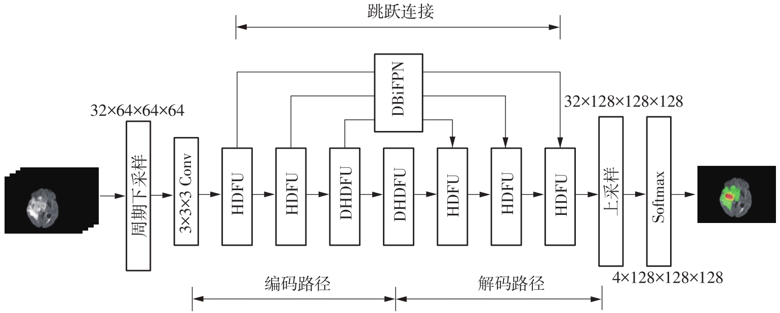
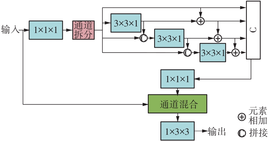
Dice similarity coefficient and Hausdorff distance under different network structures"
| 网络结构 | DD,ET | DD,WT | DD,TC | DH,ET | DH,WT | DH,TC |
|---|---|---|---|---|---|---|
| HDC | 0.773 6 | 0.893 0 | 0.818 3 | 4.188 0 | 6.744 1 | 7.977 2 |
| HDFU | 0.771 9 | 0.893 8 | 0.818 7 | 3.764 4 | 7.359 6 | 7.267 3 |
| HDFU+DHDFU | 0.775 1 | 0.893 5 | 0.824 7 | 3.890 2 | 6.351 2 | 6.438 3 |
| HDFU+DHDFU+DBiFPN | 0.777 0 | 0.900 3 | 0.830 6 | 3.874 3 | 4.864 3 | 6.691 3 |
Table 6
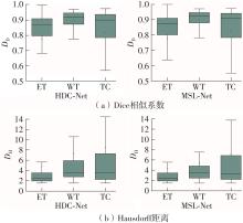
Comparison of segmentation results among seven algorithms on BraTS 2019 dataset"
| 网络模型 | Dice相似系数 | Hausdorff距离/mm | 参数量/106 | 计算量/109 | ||||
|---|---|---|---|---|---|---|---|---|
| ET | WT | TC | ET | WT | TC | |||
| 3D U-Net | 0.708 6 | 0.873 8 | 0.724 8 | 5.062 0 | 9.432 0 | 8.719 0 | 13.08 | 233.36 |
| KiU-Net | 0.732 1 | 0.876 0 | 0.739 2 | 6.323 0 | 8.942 0 | 9.893 0 | 0.29 | |
| 注意力U-Net | 0.759 6 | 0.888 1 | 0.772 0 | 5.202 0 | 7.756 0 | 8.258 0 | 34.90 | 51.30 |
| 多步级联Net | 0.771 0 | 0.886 0 | 0.813 0 | 6.033 0 | 6.232 0 | 7.409 0 | ||
| DMFNet | 0.773 3 | 0.897 5 | 0.816 7 | 2.840 0 | 6.190 0 | 6.590 0 | 3.88 | 27.04 |
| HDC-Net | 0.773 6 | 0.893 0 | 0.818 3 | 4.188 0 | 6.744 1 | 7.977 2 | 0.29 | 24.00 |
| MSL-Net | 0.777 0 | 0.900 3 | 0.830 6 | 3.874 3 | 4.864 3 | 6.691 3 | 0.39 | 31.60 |
| 1 | TABATABAI G, STUPP R, van den BENT M J,et al .Molecular diagnostics of gliomas:the clinical perspective[J].Acta Neuropathologica,2010,120(5):585-592. |
| 2 | WADHWA A, BHARDWAJ A, VERMA V S .A review on brain tumor segmentation of MRI images[J].Magnetic Resonance Imaging,2019,61:247-259. |
| 3 | CUI S, MAO L, JIANG J,et al .Automatic semantic segmentation of brain gliomas from MRI images using a deep cascaded neural network[J].Journal of Healthcare Engineering,2018,2018:4940593/1-15. |
| 4 | PINTO A, PEREIRA S, RASTEIRO D,et al .Hierarchical brain tumor segmentation using extremely rando-mized trees[J].Pattern Recognition,2018,82:105-117. |
| 5 | DEEPAK S, AMEER P M .Brain tumor classification using deep CNN features via transfer learning[J].Computers in Biology and Medicine,2019,111:103345/1-7. |
| 6 | TIWARI A, SRIVASTAVA S, PANT M .Brain tumor segmentation and classification from magnetic resonance images:review of selected methods from 2014 to 2019[J].Pattern Recognition Letters,2020,131:244-260. |
| 7 | HAVAEI M, DAVY A, WARDE-FARLEY D,et al .Brain tumor segmentation with deep neural networks[J].Medical Image Analysis,2017,35:18-31. |
| 8 | ZHAO X, WU Y, SONG G,et al .A deep learning model integrating FCNNs and CRFs for brain tumor segmentation[J].Medical Image Analysis,2018,43:98-111. |
| 9 | LONG J, SHELHAMER E, DARRELL T .Fully convolutional networks for semantic segmentation[C]∥ Proceedings of 2015 IEEE Conference on Computer Vision and Pattern Recognition.Boston:IEEE,2015:3431-3440. |
| 10 | RONNEBERGER O, FISCHER P, BROX T .U-Net:convolutional networks for biomedical image segmentation[C]∥ Proceedings of the 18th International Confe-rence on Medical Image Computing and Computer-Assisted Intervention.Munich:Springer,2015:234-241. |
| 11 | ÇICEK Ö, ABDULKADIR A, LIENKAMP S S,et al .3D U-Net:learning dense volumetric segmentation from sparse annotation[C]∥ Proceedings of the 19th International Conference on Medical Image Computing and Computer-Assisted Intervention.Athens:Springer,2016:424-432. |
| 12 | HUANG C, HAN H, YAO Q,et al .3D U2-Net:a 3D universal U-Net for multi-domain medical image segmentation[C]∥ Proceedings of the 22nd International Conference on Medical Image Computing and Computer-Assisted Intervention.Shenzhen:Springer,2019:291-299. |
| 13 | CHEN C, LIU X, DING M,et al .3D dilated multi-fiber network for real-time brain tumor segmentation in MRI[C]∥ Proceedings of the 22nd International Conference on Medical Image Computing and Computer-Assisted Intervention.Shenzhen:Springer,2019:184-192. |
| 14 | DING Y, GONG L, ZHANG M,et al .A multi-path adaptive fusion network for multimodal brain tumor segmentation[J].Neurocomputing,2020,412:19-30. |
| 15 | HUANG G, LIU Z, van der MAATEN L,et al .Densely connected convolutional networks[C]∥ Proceedings of 2017 IEEE Conference on Computer Vision and Pattern Recognition.Honolulu:IEEE,2017:4700-4708. |
| 16 | LUO Z, JIA Z, YUAN Z,et al .HDC-Net:hierarchical decoupled convolution network for brain tumor segmentation[J].IEEE Journal of Biomedical and Health Informatics,2020,25(3):737-745. |
| 17 | OKTAY O, SCHLEMPER J, FOLGOC L L,et al .Attention U-Net:learning where to look for the pancreas[EB/OL]. (2018-05-20)[2021-12-30].. |
| 18 | IBTEHAZ N, RAHMAN M S .MultiResUNet:rethin-king the U-Net architecture for multimodal biomedical image segmentation[J].Neural Networks,2020,121:74-87. |
| 19 | WANG Y L, ZHAO Z J, HU S Y,et al .CLCU-Net:cross-level connected U-shaped network with selective feature aggregation attention module for brain tumor segmentation[J].Computer Methods and Programs in Biomedicine,2021,207:106154/1-9. |
| 20 | LI X, LUO G, WANG K .Multi-step cascaded networks for brain tumor segmentation[C]∥ Proceedings of the 5th International Workshop on Brainlesion:Glioma,Multiple Sclerosis,Stroke and Traumatic Brain Injuries.Shenzhen:Springer,2019:163-173. |
| 21 | VALANARASU J M J, SINDAGI V A,HACIHALILOG- LU I,et al .KiU-Net:overcomplete convolutional architectures for biomedical image and volumetric segmentation[EB/OL]. (2021-10-14)[2021-12-30].. |
| 22 | SUDRE C H, LI W, VERCAUTEREN T,et al .Gene-ralised dice overlap as a deep learning loss function for highly unbalanced segmentations[M]∥ Deep learning in medical image analysis and multimodal learning for clinical decision support.Berlin:Springer,2017:240-248. |
| 23 | LIN T Y, GOYAL P, GIRSHICK R,et al .Focal loss for dense object detection[C]∥ Proceedings of 2017 IEEE International Conference on Computer Vision.Venice:IEEE,2017:2980-2988. |
| 24 | YU F, KOLTUN V, FUNKHOUSER T .Dilated residual networks[C]∥ Proceedings of 2017 IEEE Conference on Computer Vision and Pattern Recognition.Honolulu:IEEE,2017:472-480. |
| 25 | MEHTA S, RASTEGARI M, CASPI A,et al .ESPNet:efficient spatial pyramid of dilated convolutions for semantic segmentation[C]∥ Proceedings of the 15th European Conference on Computer Vision.Munich:Springer,2018:552-568. |
| 26 | ZHANG X, ZHOU X, LIN M,et al .ShuffleNet:an extremely efficient convolutional neural network for mobile devices[C]∥ Proceedings of 2018 IEEE Conference on Computer Vision and Pattern Recognition.Salt Lake City:IEEE,2018:6848-6856. |
| 27 | TAN M, PANG R, LE Q V .EfficientDet:scalable and efficient object detection[C]∥ Proceedings of 2020 IEEE/CVF Conference on Computer Vision and Pattern Recognition.Seattle:IEEE,2020:10781-10790. |
| [1] | TIAN Sheng, SONG Lin, ZHAO Kailong. Point Cloud Classification Based on Offset Attention Mechanism and Multi-Feature Fusion [J]. Journal of South China University of Technology(Natural Science Edition), 2024, 52(1): 100-109. |
| [2] | LI Haiyan, YIN Haolin, LI Peng, et al.. Image Inpainting Algorithm Based on Dense Feature Reasoning and Mix Loss Function [J]. Journal of South China University of Technology(Natural Science Edition), 2023, 51(9): 99-109. |
| [3] | LI Jiachun, LI Bowen, LIN Weiwei. AdfNet: An Adaptive Deep Forgery Detection Network Based on Diverse Features [J]. Journal of South China University of Technology(Natural Science Edition), 2023, 51(9): 82-89. |
| [4] | ZHAO Rongchao, WU Baili, CHEN Zhuyun, WEN Kairu, ZHANG Shaohui, LI Weihua. Graph Neural Network for Fault Diagnosis with Multi-Scale Time-Spatial Information Fusion Mechanism [J]. Journal of South China University of Technology(Natural Science Edition), 2023, 51(12): 42-52. |
| [5] | SUN Xiaohe, SHI Chenghua, LIU Linghui, et al. Concrete Crack Image Recognition System Based on Improved Seed Filling Algorithm [J]. Journal of South China University of Technology(Natural Science Edition), 2022, 50(5): 127-136,146. |
| [6] | HUANG Huaiwei, HE Wanli, CAO Yajun. Energy Harvesting Characteristics of Preloaded Piezoelectric Beams Based on Multi-Scale Approach [J]. Journal of South China University of Technology(Natural Science Edition), 2022, 50(5): 118-126. |
| [7] | YANG Chunling YANG Yajing. A Deep Neural Network Based on Layer-by-Layer Fusion of Multi-Scale Features for No-Reference Image Quality Assessment [J]. Journal of South China University of Technology(Natural Science Edition), 2022, 50(4): 81-89,141. |
| [8] | MA Li XU Mengcong ZHOU Lei. Realization of Point Line Feature Fusion PAL-SLAM Based on RGB-D Camera [J]. Journal of South China University of Technology(Natural Science Edition), 2022, 50(2): 76-83. |
| [9] | YI Qingming, LÜ Renyi, SHI Min, et al. Lightweight Object Detection Combined with Multi-Scale Dilated-Convolution and Multi-Scale Deconvolution [J]. Journal of South China University of Technology(Natural Science Edition), 2022, 50(12): 41-48. |
| [10] | HUANG Yuezhen, WANG Naizhou, LIANG Tiancai, et al. Vehicle Recognition Method Based on Improved CenterNet [J]. Journal of South China University of Technology (Natural Science Edition), 2021, 49(7): 94-102. |
| [11] | ZHANG Ruifeng, REN Guoming, LI Qiang, et al. Stereo Matching Network Based on Multi-Stage Fusion and Recurrent Aggregation [J]. Journal of South China University of Technology (Natural Science Edition), 2021, 49(6): 77-87,99. |
| [12] | YANG Yi, TAN Jiancheng, JIN Bochong, et al. Multi-Scale Simulation on the Wind Field for Complex Terrain Based on Coupled WRF and CFD Techniques [J]. Journal of South China University of Technology (Natural Science Edition), 2021, 49(5): 65-73,83. |
| [13] | CHEN Ling, DU Anlin, TANG Yukuo, et al. Effects of Structural Evolution of Starch Materials with Different Gelatinization Degrees on Materials' Printability of Hot-Extrusion 3D Printing [J]. Journal of South China University of Technology(Natural Science Edition), 2021, 49(3): 62-70,87. |
| [14] | Qi LIU Bin Yu. Pavement Crack Recognition Algorithm Based on Transposed CNN [J]. Journal of South China University of Technology(Natural Science Edition), 2021, 49(12): 124-132. |
| [15] | WANG Tichun HUA Yang QIN Jiaqi. Reasoning Model for Mechanical Product Scheme Design Based on Extensional Ontology Related Network [J]. Journal of South China University of Technology (Natural Science Edition), 2020, 48(9): 61-70. |
| Viewed | ||||||
|
Full text |
|
|||||
|
Abstract |
|
|||||

