华南理工大学学报(自然科学版) ›› 2026, Vol. 54 ›› Issue (3): 135-147.doi: 10.12141/j.issn.1000-565X.250168
X射线计算机断层扫描技术在先进材料前沿研究中的应用
崔洁1, 桂艳2, 张成毅1, 杨贤锋1
- 1.华南理工大学 分析测试中心,广东 广州 510640
2.广州职业技术大学 智能制造学院,广东 广州 511483
-
收稿日期:2025-06-06出版日期:2026-03-25发布日期:2025-09-05 -
通信作者:杨贤锋(1978—),男,博士,教授级高级工程师,主要从事无机微纳结构材料的构效关系及与此相关的仪器功能开发和新方法研究。 E-mail:czxfyang@scut.edu.cn -
作者简介:崔洁(1986 —),女,博士,正高级实验师,主要从事新能源材料构效关系及表征技术开发研究。E-mail: czcuijie@scut.edu.cn -
基金资助:国家重点研发计划项目(2022YFF0607805)
Application of X-Ray Computed Tomography in Frontier Research of Advanced Materials
CUI Jie1, GUI Yan2, ZHANG Chengyi1, YANG Xianfeng1
- 1.Analytical and Testing Center of SCUT,South China University of Technology,Guangzhou 510640,Guangdong,China
2.School of Intelligent Manufacturing,Guangzhou Polytechnic University,Guangzhou 511483,Guangdong,China
-
Received:2025-06-06Online:2026-03-25Published:2025-09-05 -
Contact:杨贤锋(1978—),男,博士,教授级高级工程师,主要从事无机微纳结构材料的构效关系及与此相关的仪器功能开发和新方法研究。 E-mail:czxfyang@scut.edu.cn -
About author:崔洁(1986 —),女,博士,正高级实验师,主要从事新能源材料构效关系及表征技术开发研究。E-mail: czcuijie@scut.edu.cn -
Supported by:the National Key R & D Program of China(2022YFF0607805)
摘要:
作为一种先进的无损三维成像检测技术,X射线计算机断层扫描(CT)可实现样品内部结构的可视化表征。该技术基于X射线与物质的相互作用机制,采集X射线穿透样品后的信号成像,再以计算机算法处理获取的断层图像,最终实现对样品的三维重构。凭借高密度分辨率、便捷的数字化处理等优势,该技术已在医学诊断、工业检测等领域取得重大突破。该文重点围绕X射线CT技术在以结构材料和新能源材料为代表的先进材料领域中的应用展开研究,系统梳理其基于X射线衰减、信号转换与三维重构的核心原理;聚焦材料科学应用,通过航空航天构件焊缝缺陷检测、电子封装焊点隐形缺陷识别、增材制造材料孔隙量化等实例,阐明CT技术在缺陷定位、损伤追踪、微观结构量化中的作用;借助该技术在锂电池电极演化、燃料电池水管理、金属负极枝晶追踪等研究中的应用,凸显其在揭示材料结构与电化学性能的关联、器件设计优化、安全性提升等方面的作用;同时,总结其“无损 + 三维定量 + 动态追踪”等优势,剖析纳米级成像效率低、数据融合难等瓶颈,并从新型探测器研发、人工智能(AI)辅助重建、多技术联用等维度,展望未来突破路径,为其在结构材料性能提升、新能源材料研发中的深度应用提供方向。这些探讨可为科研人员提供技术创新方向,有助于提升我国高端检测设备的自主研发能力。
中图分类号:
引用本文
崔洁, 桂艳, 张成毅, 杨贤锋. X射线计算机断层扫描技术在先进材料前沿研究中的应用[J]. 华南理工大学学报(自然科学版), 2026, 54(3): 135-147.
CUI Jie, GUI Yan, ZHANG Chengyi, YANG Xianfeng. Application of X-Ray Computed Tomography in Frontier Research of Advanced Materials[J]. Journal of South China University of Technology(Natural Science Edition), 2026, 54(3): 135-147.

图1
Nano-CT和Micro-CT工作原理图"
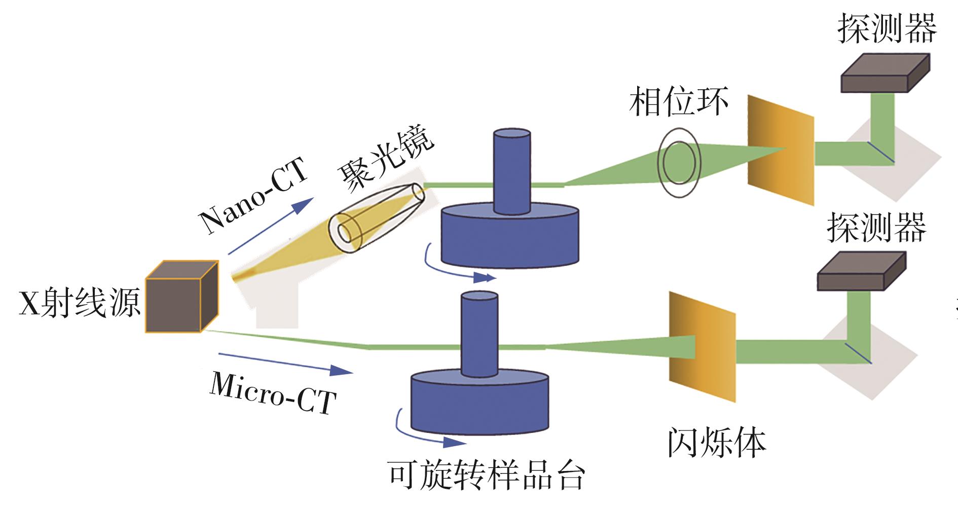

图2
当X射线束穿过0.5 mm铜、3 mm铝或30 mm软组织时产生等效强度的示例[11]"
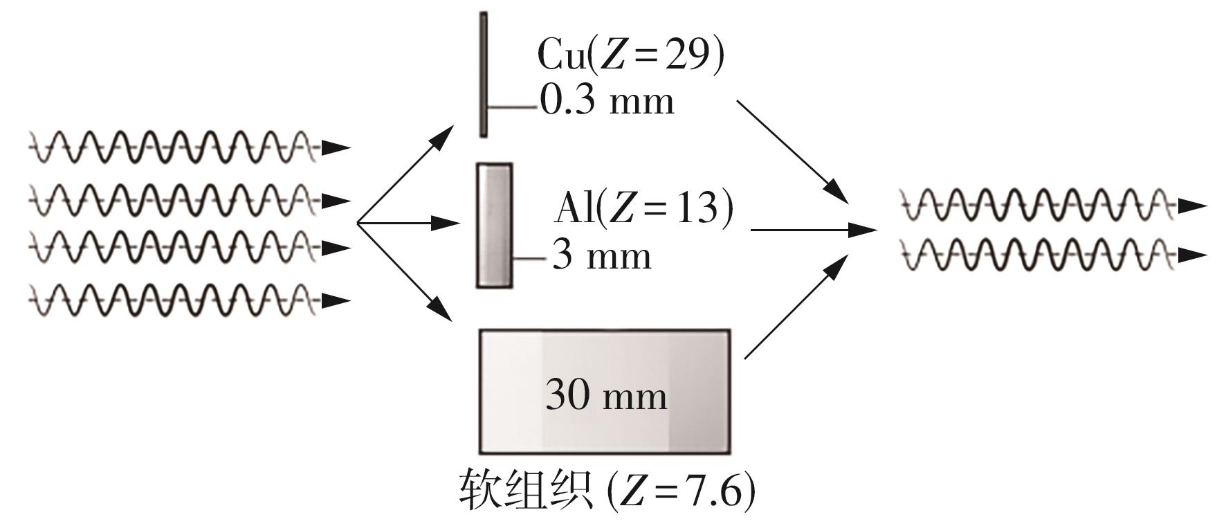
图3
商业铝材摩擦焊接样品中内部缺陷的功率/力信号的定位分析及X射线CT检测[22]"
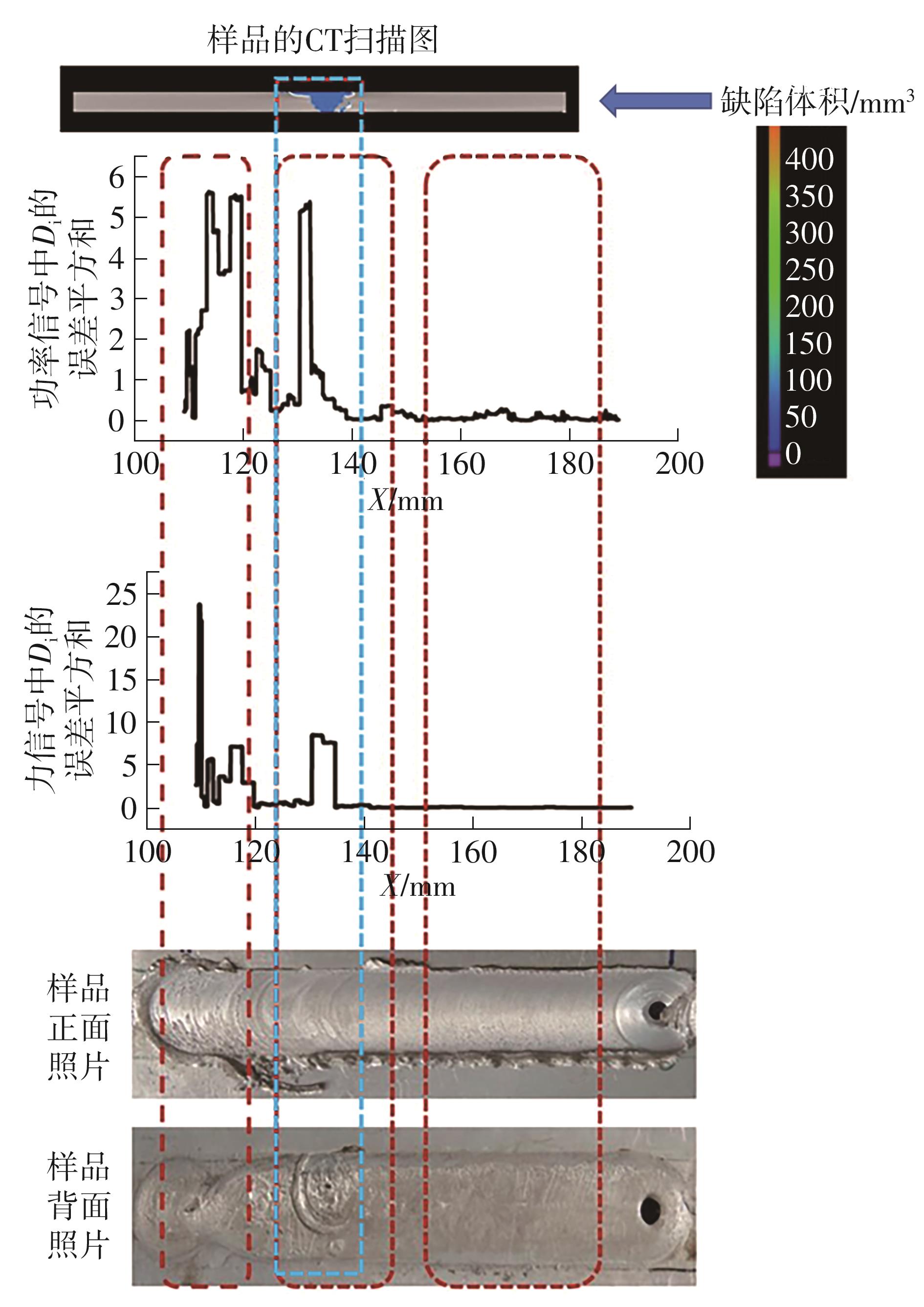
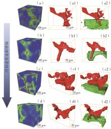
图4
GW63K合金微观凝固组织的三维形貌CT结果[28]"
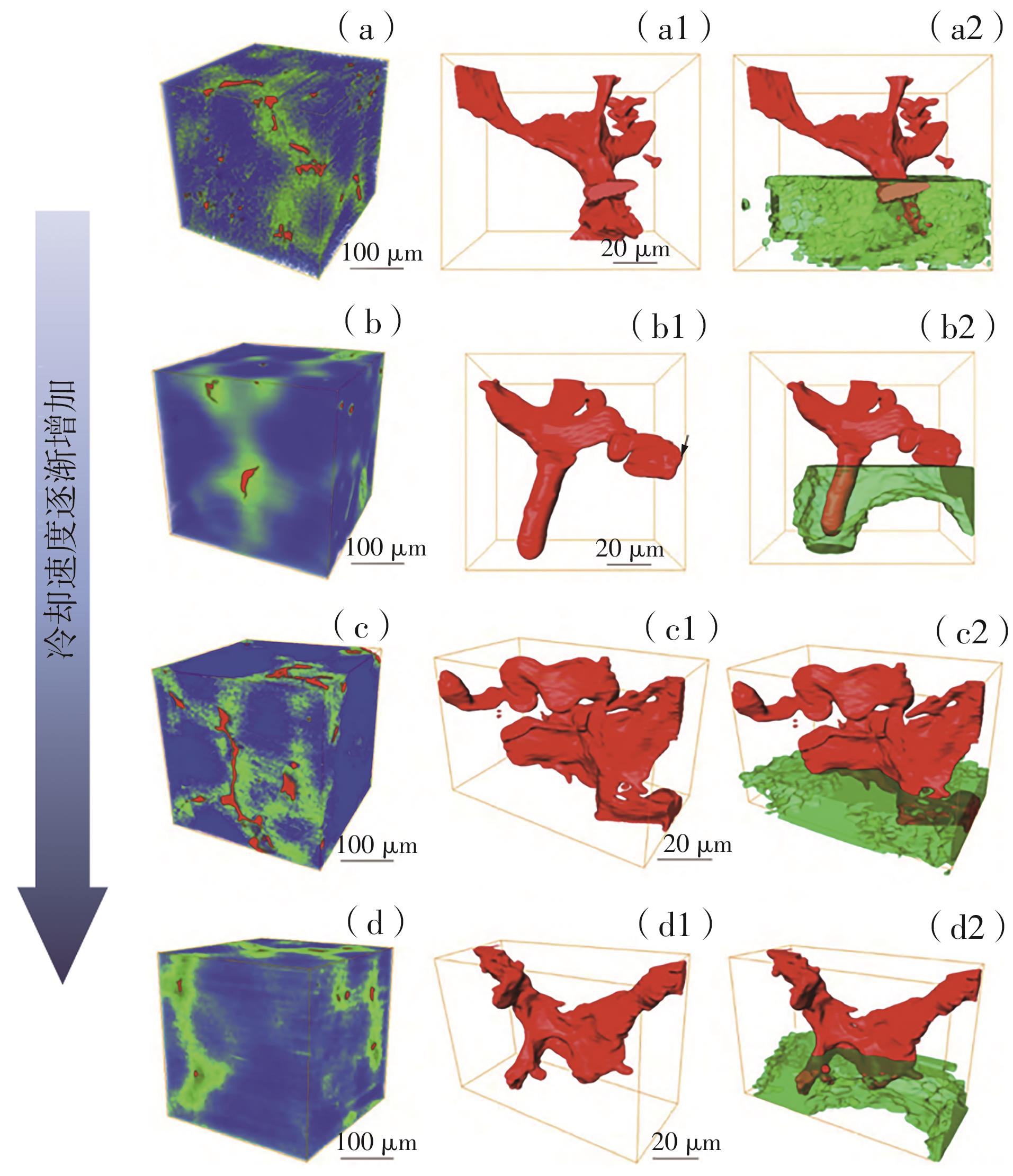

图5
负载过渡金属的氮掺杂碳基催化剂热解动态演化的原位变温Micro-CT研究[37]"
图6
X射线CT研究燃料电池催化剂层内的水分布情况[44]"
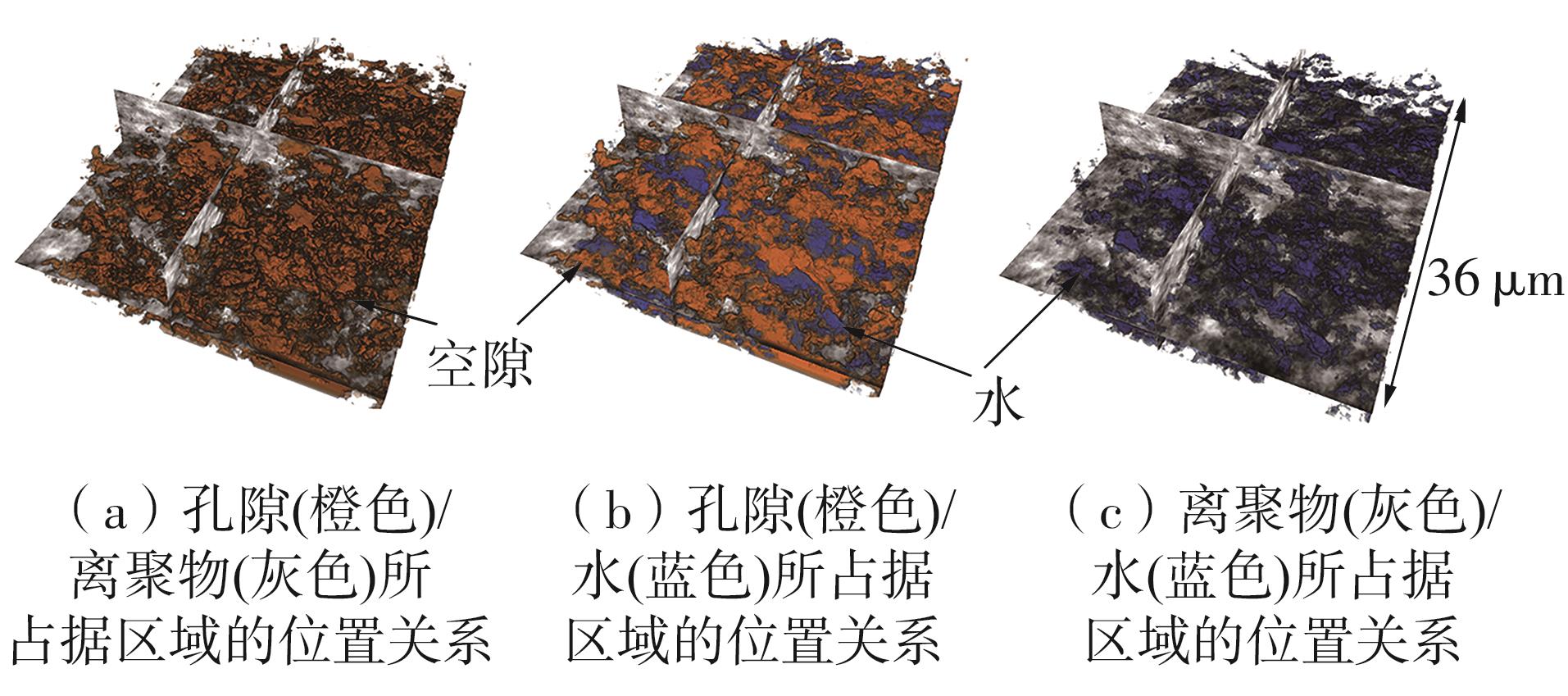
图7
Li0.5Ni0.6Mn0.2Co0.2O2二次颗粒加热前后元素化学价态演化的全X射线光谱显微成像分析[56]"
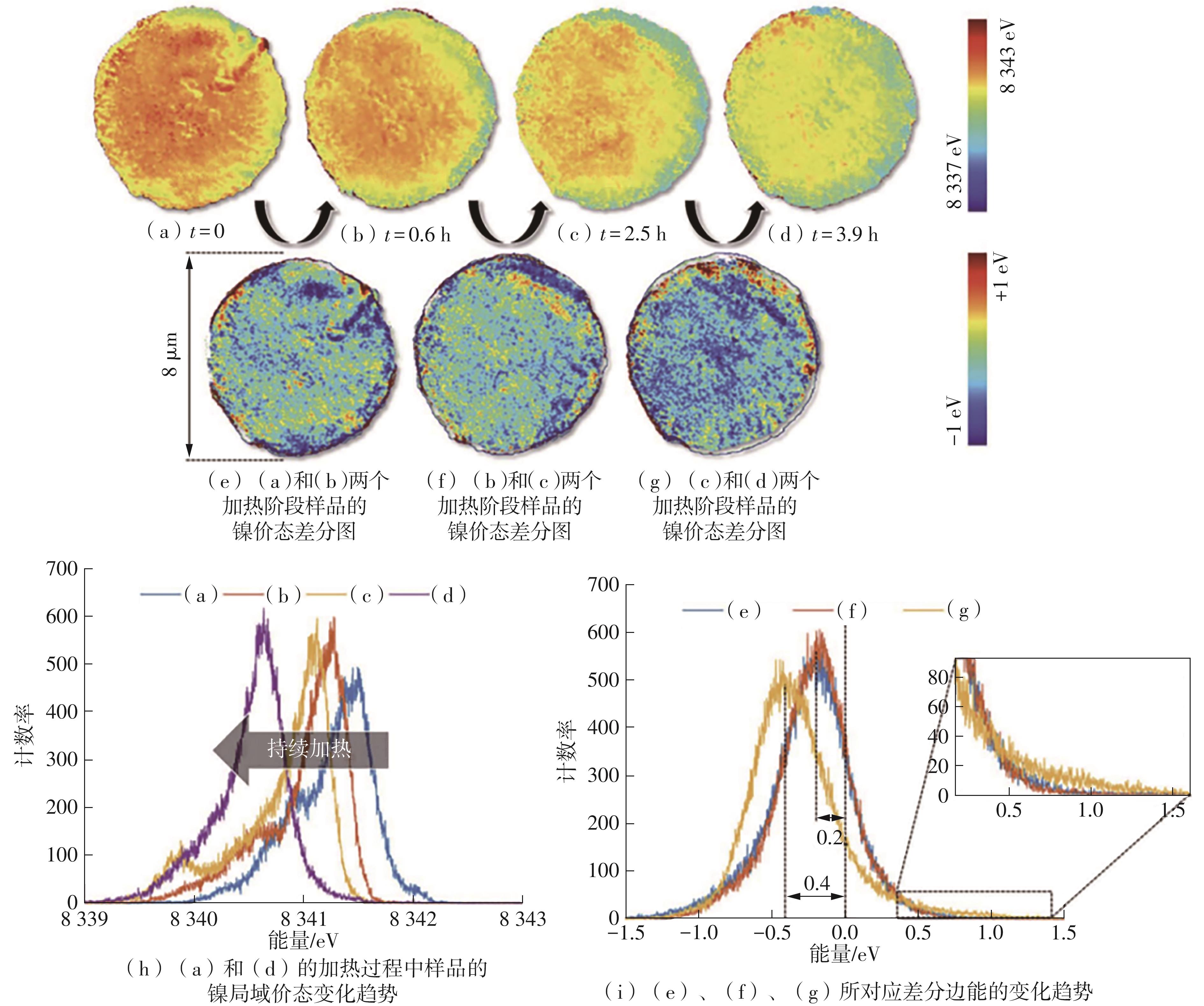
图8
重复镀-剥-镀过程中锌沉积的原位X射线CT扫描和软X射线吸收光谱分析[64]"
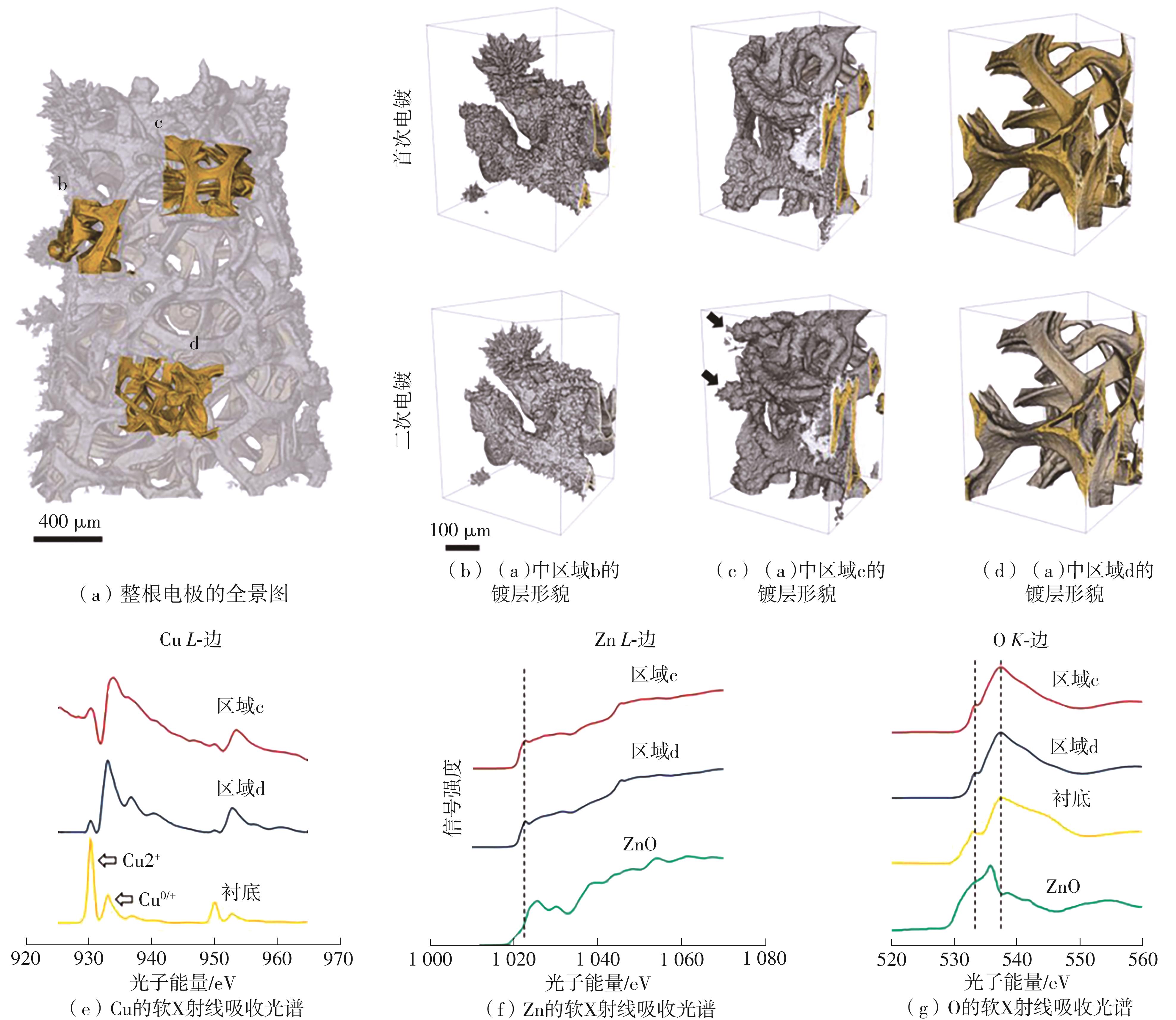
表1
X射线CT技术在材料研究领域应用中的问题分析研究"
| 技术瓶颈 | 具体表现 | 典型案例 | |
|---|---|---|---|
| 结构材料表征领域 | 宏观覆盖与微观精度的矛盾 | 大型构件需厘米级视场保证完整性,但Micro-CT在此尺度下分辨率降低,难以识别微小缺陷 | 航天器燃料箱焊缝等大型构件需厘米级视场,Micro-CT在此尺度下分辨率降至5 μm以上,无法识别小于1 μm的焊缝间隙[ |
| 动态损伤演化捕捉能力不足 | 同步辐射CT虽能追踪疲劳裂纹扩展,但时间分辨率不够,无法捕捉瞬时断裂过程 | 现有1 Hz的时间分辨率无法捕捉冲击载荷下的毫秒级瞬时断裂过程,导致裂纹尖端应力场分析存在时间盲区[ | |
| 高原子序数材料穿透有困难 | 金属材料高原子序数特性导致X射线穿透深度不足,需增加曝光时间,还可能引入伪影 | 对厚度> 5 mm的Ti64合金构件,需增加曝光时间3倍,且会因样品台漂移引入伪影[ | |
| 新能源材料表征领域 | 轻元素成像精度低 | 轻元素体系依赖相位衬度成像,但受同步辐射装置机时限制,实验室级相位衬度CT识别率低 | 实验室级相位衬度CT的硫相识别率仅65%,而同步辐射的识别率可以达92%[ |
| 原位测试稳定性差 | 电解液挥发导致样品漂移,电极膨胀,使枝晶生长轨迹三维重建误差大 | 样品漂移> 2 μm,电极循环过程中厚度变化达10%时枝晶生长轨迹的重建误差超15%[ | |
| 高温环境干扰较大 | 高温下样品台热漂移易导致孔隙率计算偏差,难以建立精准关联 | 800 ℃高温下样品台热漂移会导致孔隙率计算偏差达8%[ | |
| 跨领域的共性问题 | 数据处理效率不足 | 大型数据集重建耗时过长,超出传统设备处理能力 | 增材制造构件全尺寸扫描数据(10 TB)需72 h重建;电池动态数据集(1 TB/h)难以实时处理[ |
| 辐射损伤的材料存在差异性 | 不同材料对辐射剂量的耐受度矛盾 | 聚合物粘结剂在10⁴Gy剂量下分子链断裂率> 30%,但金属材料需更高剂量才能保证衬度[ | |
| 多模态数据融合技术有断层 | 结构材料与新能源材料的“结构-性能”模型独立,缺乏统一分析框架 | 结构材料“CT-力学性能”模型与新能源材料“CT-电化学性能”模型无法复用,制约技术普适性[ | |
| [1] | NICHOLLS M .Sir Godfrey Newbold Hounsfield and Allan M.Cormack[J].European Heart Journal,2019,40(26):2101-2103. |
| [2] | STEINBOCK L, DUSTMANN C H .Investigation of the inner structures of zebra cells with a microtomograph [J].Journal of The Electrochemical Society,2001,148(1):A132-A136. |
| [3] | CARTER R, HUHMAN B, LOVE C T,et al .X-ray computed tomography comparison of individual and parallel assembled commercial lithium iron phosphate batteries at end of life after high-rate cycling[J].Journal of Power Sources,2018,381:46-55. |
| [4] | LOVERIDGE M, REMY G, KOURRA K,et al .A looking deeper into the galaxy (note 7)[J].Batteries,2018,4(1):3/1-11. |
| [5] | SALVO L, CLOETENS P, MAIRE E,et al .X-ray micro-tomography an attractive characterization technique in materials science[J].Nuclear Instruments and Methods in Physics Research,Section B:Beam Interactions with Materials and Atoms,2003,200:273-286. |
| [6] | VILLARRAGA-GÓMEZ H, HERAZO E L, SMITH S T .X-ray computed tomography:from medical imaging to dimensional metrology[J].Precision Engineering,2019,60:544-569. |
| [7] | WITHERS P J, BOUMAN C, CARMIGNATO S,et al .X-ray computed tomography[J].Nature Reviews Me-thods Primers,2021,1(18):1-21. |
| [8] | WANG G, YU H, DE MAN B .An outlook on X-ray CT research and development[J].Medical Physics,2008,35(3):1051-1064. |
| [9] | MAIRE E, WITHERS P J .Quantitative X-ray tomography[J].International Materials Reviews,2014,59(1):1-43. |
| [10] | HEENAN T M M, TAN C, HACK J,et al .Developments in X-ray tomography characterization for electrochemical devices[J].Materials Today,2019,31:69-85. |
| [11] | HUDA W, ABRAHAMS R B .Radiographic techniques,contrast,and noise in X-ray imaging[J].American Journal of Roentgenology,2015,204(1):W126-W131. |
| [12] | ZIELKE L, BARCHASZ C, WALUŚ S,et al .Degradation of Li/S battery electrodes on 3D current collectors studied using X-ray phase contrast tomography[J].Scientific Reports,2015,5:10921/1-12. |
| [13] | BÜHRER M, STAMPANONI M, ROCHET X,et al .High-numerical-aperture macroscope optics for time-resolved experiments[J].Journal of Synchrotron Radiation,2019,26(5):1161-1172. |
| [14] | XU H, MARONE F, NAGASHIMA S,et al .Exploring sub-second and sub-micron X-ray tomographic ima-ging of liquid water in PEFC gas diffusion layers[J].ECS Transactions,2019,92(8):11-21. |
| [15] | ELLER J, ROTH J, MARONE F,et al .Towards ultra-fast X-ray tomographic microscopy of liquid water in PEFC[J].ECS Transactions,2011,41(1):387-394. |
| [16] | KULKARNI D, HUYNH A, SATJARITANUN P,et al .Elucidating effects of catalyst loadings and porous transport layer morphologies on operation of proton exchange membrane water electrolyzers[J].Applied Catalysis B:Environmental,2022,308:121213/1-15. |
| [17] | MIAO N,HAI B, WANG S F,et al .An in-situ X-ray computed tomography imaging apparatus with stack pressures for rechargeable batteries[J].Scripta Materialia,2023,229:115381/1-5. |
| [18] | ROTH J, ELLER J, BÜCHI F N .Effects of synchrotron radiation on fuel cell materials[J].Journal of the Electrochemical Society,2012,159(8):F449-F455. |
| [19] | WANG J, MORIN C, LI L,et al .Radiation damage in soft X-ray microscopy[J].Journal of Electron Spectroscopy and Related Phenomena,2009,170(1/2/3):25-36. |
| [20] | HOWELLS M R, BEETZ T, CHAPMAN H N,et al .An assessment of the resolution limitation due to radiation damage in X-ray diffraction microscopy[J].Journal of Electron Spectroscopy and Related Phenomena,2009,170:4-12. |
| [21] | SCHNEIDER A, WIESER C, ROTH J,et al .Impact of synchrotron radiation on fuel cell operation in imaging experiments[J].Journal of Power Sources,2010,195(19):6349-6355. |
| [22] | ROY R B, GHOSH A, BHATTACHARYYA S,et al .Weld defect identification in friction stir welding through optimized wavelet transformation of signals and validation through X-ray micro-CT scan[J].The International Journal of Advanced Manufacturing Techno-logy,2018,99(1/2/3/4):623-633. |
| [23] | SHALOO M, SCHNALL M, KLEIN T,et al .A review of nondestructive testing (NDT) techniques for defect detection:application to fusion welding and future wire arc additive manufacturing processes[J].Materials,2022,15(3697):1-26. |
| [24] | LEE M C, CHEN W T, LIN C T,et al .Detection of micro defects in 3DIC packages by means of non-destructive 3D X-ray[C]∥ Proceedings of the 2012 7th International Microsystems,Packaging,Assembly and Circuits Technology Conference (IMPACT).Taipei:IEEE CPMT-Taiwan,IMAPS-Taiwan,Industrial Technology Research Institute (ITRI),Taiwan Printed Circuit Association(TPCA),2012:209-211. |
| [25] | KE C Y, CHEN L P .X-ray inspection for fomcm (fan-out multi chip module) μ-bump non-wet[C]∥ Proceedings of 2021 IEEE 23rd Electronics Packaging Technology Conference (EPTC).Singapore:IEEE,2021:93-94. |
| [26] | 施国栋,何德坪,张勇明,等 .超轻多孔金属孔结构的X射线断层扫描分析[J].机械工程材料,2008,32(3):13-15. |
| SHI Guo-dong, HE De-ping, ZHANG Yong-ming,et al .Analysis of pore structure for ultra-light porous metals by X-ray tomography[J].Materials for Mechanical Engineering,2008,32(3):13-15. | |
| [27] | 张臻 .基于X射线测量的铝合金铸件微观缺陷预测及分析[J].铸造技术,2014,35(11):2760-2762. |
| ZHANG Zhen .Prediction and analysis on micro defect in aluminum alloy castings based on X-ray measurement [J].Foundry Technology,2014,35(11):2760-2763. | |
| [28] | 赵雪婷,张天向,李少翔,等 .Mg-Gd-Y-Zr合金可控冷却速率实验方法及凝固组织的X射线断层扫描表征[J].中国有色金属学报,2022,32(7):1911-1923. |
| ZHAO Xue-ting, ZHANG Tian-xiang, LI Shao-xiang,et al .Preparation of Mg-Gd-Y-Zr alloy solidified at controlled cooling rate and microstructure characterization by X-ray tomograph[J].The Chinese Journal of Nonferrous Metals,2022,32(7):1911-1923. | |
| [29] | BUFFIÈRE J Y, CLOETENS P, LUDWIG W,et al .In situ X-ray tomography studies of microstructural evolution combined with 3D modeling[J].MRS Bulletin,2008,33(6):611-619. |
| [30] | ZENYUK I V .Bridging X-ray computed tomography and computational modeling for electrochemical energy conversion and storage[J].Current Opinion in Electrochemistry,2019,13:78-85. |
| [31] | HEENAN T M M, TAN C, HACK J,et al .Developments in X-ray tomography characterization for electrochemical devices[J].Materials Today,2019,31:69-85. |
| [32] | BRAATEN J P, OGAWA S, YARLAGADDA V,et al .Studying Pt-based fuel cell electrode degradation with nanoscale X-ray computed tomography[J].Journal of Power Sources,2020,478(229049):1-7. |
| [33] | JIA Q, RAMASWAMY N, TYLUS U,et al .Spectroscopic insights into the nature of active sites in iron-nitrogen-carbon electrocatalysts for oxygen reduction in acid and the Redox mechanisms[J].Nano Energy,2016,29:65-82. |
| [34] | JIA Q, RAMASWAMY N, HAFIZ H,et al .Experimental observation of redox-induced Fe-N switching behavior as a determinant role for oxygen reduction activity[J].ACS Nano,2015,9(12):12496-12505. |
| [35] | BARNARD H S, MACDOWELL A, PARKINSON D Y,et al .Synchrotron X-ray micro tomography at the advanced light source:in-situ sample environments for advanced aerospace materials[J].Microscopy and Microanalysis,2018,24(S2):444-445. |
| [36] | LITSTER S, HESS K, EPTING W,et al .Catalyst layer analysis:nanoscale X-ray CT,spatially-resolved in situ microscale diagnostics and modeling[J].ECS Transactions,2011,41(4):409-418. |
| [37] | HUANG Y, CHEN Y, XU M,et al .Catalysts by pyrolysis:direct observation of chemical and morphological transformations leading to transition metal-nitrogen-carbon materials[J].Materials Today,2021,47:53-68. |
| [38] | CHEN Y, HUANG Y, XU M,et al .Catalysts by pyrolysis:direct observation of transformations during re-pyrolysis of transition metal-nitrogen-carbon materials leading to state-of-the-art platinum group metal-free electrocatalyst[J].Materials Today,2022,53:58-70. |
| [39] | LIU S, LI C, ZACHMAN M J,et al .Atomically dispersed iron sites with a nitrogen-carbon coating as highly active and durable oxygen reduction catalysts for fuel cells[J].Nature Energy,2022,7:652-663. |
| [40] | LI J, SOUGRATI M T, ZITOLO A,et al .Identification of durable and non-durable FeN x sites in Fe-N-C materials for proton exchange membrane fuel cells[J].Nature Catalysis,2021,4:10-19. |
| [41] | GRUNWALDT J D, KIMMERLE B, BAIKER A,et al .Catalysts at work:from integral to spatially resolved X-ray absorption spectroscopy[J].Catalysis Today,2009,145(3):267-278. |
| [42] | WEBER A Z, BORUP R L, DARLING R M,et al .A critical review of modeling transport phenomena in polymer-electrolyte fuel cells[J].Journal of The Electrochemical Society,2014,161(12):F1254-F1299. |
| [43] | NOVÁK V, DUDÁK M, KOČÍ P,et al .Understanding the gas transport in porous catalyst layers by using digital reconstruction techniques[J].Current Opinion in Chemical Engineering,2015,9:16-26. |
| [44] | NORMILE S J, SABARIRAJAN D C, CALZADA O,et al .Direct observations of liquid water formation at nano-and micro-scale in platinum group metal-free electrodes by operando X-ray computed tomography[J].Materials Today Energy,2018,9:187-197. |
| [45] | MOSS A B, HÄTINEN J, KÚŠ P,et al .Versatile high energy X-ray transparent electrolysis cell for operando measurements[J].Journal of Power Sources,2023,562:232754/1-19. |
| [46] | KATO S, YAMAGUCHI S, YOSHIMUNE W,et al.Ex-situ visualization of the wet domain in the microporous layer in a polymer electrolyte fuel cell by X-ray computed tomography under water vapor supply[J].Electrochemistry Communications,2020,111:106644/1-5. |
| [47] | QIAN J, HENDERSON W A, XU W,et al .High rate and stable cycling of lithium metal anode[J].Nature Communications,2015,6:6362/1-9. |
| [48] | STEPHENSON D E, WALKER B C, SKELTON C B,et al .Modeling 3D microstructure and ion transport in porous Li-ion battery electrodes[J].Journal of the Electrochemical Society,2011,158(7):A781-A789. |
| [49] | XU Y, HU E, ZHANG K,et al .In situ visualization of state-of-charge heterogeneity within a LiCoO₂ particle that evolves upon cycling at different rates[J].ACS Energy Letters,2017,2(5):1240-1245. |
| [50] | YU X, FENG Z, REN Y,et al .Simultaneous ope-rando measurements of the local temperature,state of charge,and strain inside a commercial lithium-ion battery pouch cell[J].Journal of the Electrochemical Society,2018,165(7):A1578-A1585. |
| [51] | CHEN G, SONG X, RICHARDSON T J .Electron microscopy study of the LiFePO₄ to FePO₄ phase transition [J].Electrochemical and Solid-State Letters,2006,9(6):A295-A298. |
| [52] | SHIM J, STRIEBEL K A .Effect of electrode density on cycle performance and irreversible capacity loss for natural graphite anode in lithium-ion batteries[J].Journal of Power Sources,2003,119/120/121:934-937. |
| [53] | MÜLLER S, ELLER J, EBNER M,et al .Quantifying inhomogeneity of lithium-ion battery electrodes and its influence on electrochemical performance[J].Journal of the Electrochemical Society,2018,165(2):A339-A344. |
| [54] | LU X, BERTEI A, FINEGAN D P,et al .3D microstructure design of lithium-ion battery electrodes assisted by X-ray nano-computed tomography and modelling[J].Nature Communications,2020,11:2079/1-13. |
| [55] | TIAN C, XU Y, NORDLUND D,et al .Charge he-terogeneity and surface chemistry in polycrystalline ca-thode materials[J].Joule,2018,2(3):464-477. |
| [56] | WEI C X, ZHANG Y, LEE S J,et al .Thermally driven mesoscale chemomechanical interplay in Li0.5Ni0.6Mn0.2Co0.2O2 cathode materials[J].Journal of Materials Chemistry A,2018,6:23055-23061. |
| [57] | CHENG X B, ZHANG R, ZHAO C Z,et al .Toward safe lithium metal anode in rechargeable batteries:a review[J].Chemical Reviews,2017,117(17):10403-10473. |
| [58] | ASLAM M K, NIU Y, HUSSAIN T,et al .How to avoid dendrite formation in metal batteries:innovative strategies for dendrite suppression[J].Nano Energy,2021,86:106142/1-29. |
| [59] | YANG Q, LI Q, LIU Z,et al .Dendrites in Zn-based batteries[J].Advanced Materials,2020,32:2001854/1-32. |
| [60] | DENG Z, LIN X, HUANG Z,et al .Recent progress on advanced imaging techniques for lithium-ion batteries[J].Advanced Energy Materials,2021,11:2000806/1-24. |
| [61] | ZAN G B, ZHANG J, MONACO F,et al .Understanding multi-scale battery degradation with a macro-to-nano zoom through its hierarchy[J].Journal of Mate-rials Chemistry A,2021,9(35):19886-19893. |
| [62] | HAO S, DAEMI S R, HEENAN T M M,et al .Tracking lithium penetration in solid electrolytes in 3D by in-situ synchrotron X-ray computed tomography [J].Nano Energy,2021,82:105744/1-9. |
| [63] | QIAN G N, ZAN G B, PIANETTA P,et al .Perspective—morphology and dynamics of metal dendrites in batteries revealed by X-ray computed tomography[J].Journal of the Electrochemical Society,2023,169:120540/1-5. |
| [64] | QIAN G N, ZAN G B, LI J Z,et al .Structural,dynamic, and chemical complexities in zinc anode of an operating aqueous Zn-ion battery[J].Advanced Ener-gy Materials,2022,12:2200255/1-10. |
| [1] | 叶玮琳, 涂子涵, 肖旭鹏, 等. 基于LabVIEW的TDLAS检测系统多参数影响研究[J]. 华南理工大学学报(自然科学版), 2021, 49(6): 141-148. |
| 阅读次数 | ||||||
|
全文 |
|
|||||
|
摘要 |
|
|||||